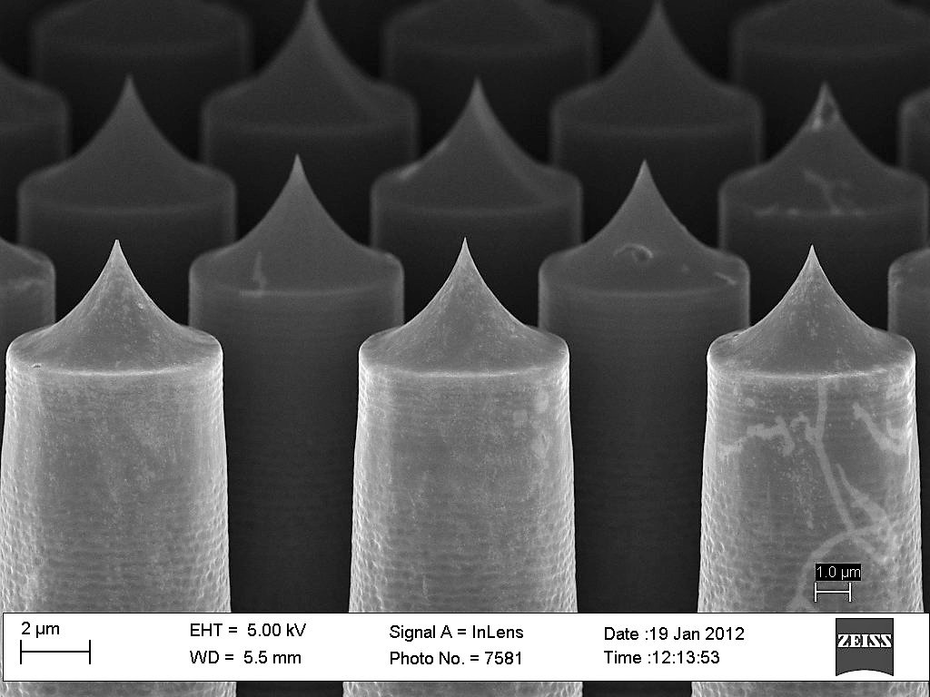
This is a scanning electron microscope image of silicon nannoneedles developed by Seungkeun Choi for drug delivery research. The sharp tip is used to puncture cells. (Photo courtesy of Seungkeun Choi)
UW Bothell researchers won $460,000 from the National Science Foundation for a scanning electron microscope that can see objects as small as one nanometer — a billionth of an inch. A piece of paper is about 100,000 nanometers thick.
Researchers plan to use the microscope for nanotechnology projects and research in biology and biotechnology.
The new microscope saves the time and expense of using a scanning microscope on the UW Seattle campus and creates more opportunities for research by UW Bothell graduate and undergraduate students said Seungkeun Choi, the principal investigator. Having it a UW Bothell also will promote and inspire science and technology opportunities through the community, Choi said.
A room is being prepared in Discovery Hall to house the microscope. The Hitachi SU5000 should be installed by the end of December and ready to use as soon as winter quarter, Choi said.
Bothell was able to present a winning grant proposal because of the multiple disciplines represented by the research team planning to use the microscope: Choi and Hung Cao in electrical engineering, John Bridge and Cassandra Wright in mechanical engineering, and Kristina Hillesland in biology.
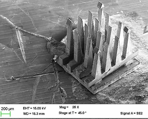
Photo: Electrode array image from a scanning electron microcsope (Courtesy of Hung Cao)
The world-class instrument will see a lot of use from various disciplines, said Elaine Scott, dean of the School of Science, Technology, Engineering & Mathematics. “We are so excited about having this high-level instrumentation that will be available to our faculty and our students,” Scott said.
Choi, an assistant professor in electrical engineering, plans to use the scanning electron microscope to develop light-trapping structures for organic solar cells.
Cao, an assistant professor in electrical engineering, plans to use the microscope to enhance extremely small biosensors, such as ones Cao uses to study heart regeneration in zebrafish, something that one day could help people who suffer heart attacks.
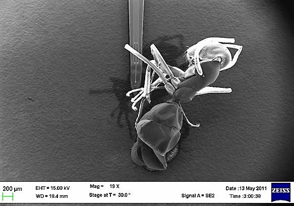
Photo: An ant under the scanning electron microscope (Courtesy of Hung Cao)
“This means a lot to our research as we use it regularly to characterize the microdevices and microstructures,” said Cao.

Hillesland, an assistant professor in biology, plans to study mutualism — beneficial interactions between species — over the course of more than 2,000 generations of bacteria.
Bridge, an associate professor of mechanical engineering, plans to study the materials used in medical devices. And Wright, an assistant professor of mechanical engineering, plans to study new materials for joint replacements to slow their wear and increase their service life.
In addition, more than 10 STEM courses will use the scanning electron microscope.